Patellar instability means the kneecap (patella) is unstable. This occurs when the patella moves out of the groove at the end of the femur, whose function is to keep the kneecap in place. When we bend or straighten the knee, the patella moves up and down along a V-shaped groove called the trochlea. In cases of patellar instability, the patella does not move correctly within the groove.
The patella is part of the extensor mechanism. Several tissues (muscles, tendons, and ligaments) that run across the front of the thigh bone (femur) pass over the patella and attach to the main bone of the lower leg (tibia). These muscles pull the patella upward through the trochlear groove when the leg is extended and downward when it is bent. If the patella is unstable, it moves out of the groove. The main causes of patellar instability include: a shallow or irregular trochlear groove; loose ligaments or overly flexible joints; direct trauma to the kneecap during a fall, sports injury, or accident.
When the patella slips out of the trochlear groove, you may feel the knee give way. You may be unable to bear weight on the affected leg or even stand. Often there is an inability to straighten the knee or walk. Other symptoms of patellar instability include: knee pain, stiffness, and swelling; clicking or popping sounds in the knee when climbing stairs or bending it; a feeling that the patella is catching or shifting from side to side. These symptoms may not be present daily and might only appear occasionally or during sports activities.
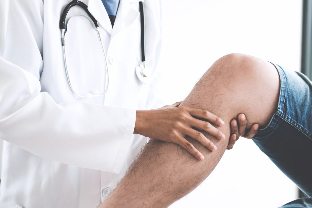
You should consult an orthopedic knee surgeon to establish a diagnosis and receive treatment advice. It is important to assess the mobility of your knee, check for significant joint swelling, and perform the patellar apprehension test to determine at what degrees of motion your patella becomes unstable. Most often, an X-ray is recommended to confirm if the patella is still out of place or has returned to its normal position and to rule out fractures. However, an MRI should be performed to assess associated injuries such as ligament or meniscal damage, or the presence of loose bodies. Sometimes, when the patella dislocates, the forces involved are so strong that fragments of cartilage and bone can detach and remain loose within the knee.
After the first episode of patellar dislocation, and if there are no loose bodies in the knee or major risk factors for instability, the recommended treatment in most cases is physiotherapy to reduce pain and swelling, and to restore mobility and muscle strength. In cases where the patella has dislocated more than once, or in certain first-time dislocations, surgery may be necessary.
Most surgeries for patellar instability involve arthroscopy, which uses small incisions and the insertion of a camera into the joint to assess cartilage damage and other associated injuries. In all cases, reconstruction of the medial patellofemoral ligament (MPFL) is necessary. This ligament acts like a rope that keeps the patella within the trochlear groove and is almost always damaged during the first dislocation. If there are abnormalities in patellar alignment, osteotomies (bone corrections) of the proximal tibia may be needed to
reposition the patella more favorably. Finally, in people with a shallow or abnormal trochlear groove, a trochleoplasty may be indicated. This involves sculpting the bone beneath the femoral cartilage to create a new groove for the patella to fit into. Our team was a pioneer in Portugal in introducing the arthroscopic trochleoplasty technique, which allows excellent results with faster recovery and less pain.
We created this concept to give it our full attention. The complexity of the knee joint requires careful assessment and differentiated knowledge.
Copyright ©2025 Knee Surgery. Privacy Policy. Powered by WEbrand Agency